Read The Root of Thought Online
Authors: Andrew Koob
The Root of Thought (6 page)
Throughout the brain, microglia (the smallest of all the glial cells) respond to injury and infection like the T cells and B cells in the blood. These cells look like ice cream sprinkles when they migrate toward an area of infection. They can be considered the Red Cross of the brain.
However, the cell that has provided such startling discoveries recently is the astrocyte (see
Figure 4.1
). The astrocytes are the cities to the neuronal highways. This cell is different functionally from the other
glial cells. The astrocyte is the most abundant cell in the human cortex and was first described by Otto Deiters (1834–1863) in an unfinished manuscript in the 1850s. Deiters died at the age of 29, and it was another former student of Johannes Müller—Max Schulte (1825–1874)—who published it for him. Deiters supported Virchow’s view that the cells were simply connective tissue. He described the flat star-shaped cells with many processes in the white and grey matter.
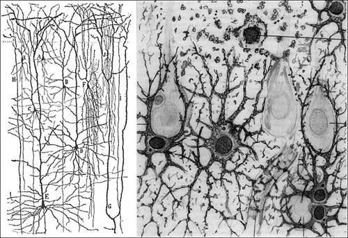
FIGURE 4.1 On the left are drawings of neurons in the cortex by Cajal. On the right is a closer view of astrocytes (dark, spidery cells) hanging out next to neuron cell bodies (pale cells with axons extending up).
Jakob Henle (1809–1885), who was also a student of Müller’s, believed that the cells were active, functional units in the brain. In 1856, Virchow wrote that Henle was blocking his concept of glia as connective tissue simply to serve “his own interests.” In 1869, Henle published his first drawing of these cells.
Later, Golgi conducted studies with his stain where he claimed glia contact with the blood vessels and neurons. Golgi’s discovery of the astrocyte’s contact with the blood vessels would be the beginning of his idea that the function of astrocytes is to provide nourishment for neurons.
Cajal was quick to discount Golgi, but he had no idea about the nature of astrocytes himself. In his struggles with glia, he once wrote, “What is the function of these glial cells in neural centers? The answer is still not known and the problem is even more serious because it might remain unsolved for many years to come until physiologists find direct methods to attack it.”
The word astrocyte sounds like an intergalactic parade, and it was used to demonstrate the star-shaped cells. Michael von Lenhossek (1863–1937) came up with the term in 1891 to describe the “neuroglia.” Neuroglia was a vogue term in the nineteenth century as the neuron was being put on the pedestal. Neuroglia meant everything except for neurons. However, they mainly referred to astrocytes as they were abundant, and oligodendricytes had not been discovered yet. Astrocytes were also previously called “spider cells.” German pathologist Edgar von Gierke (1877–1945) called the term inadequate because “nobody has seen a spider with as many feet as these cells have processes.” Lenhossek thought all glia should be termed spongicytes and divided into subsets, one of which would be astrocytes.
Some cells believed to be related to astrocytes are Bergmann glia in the cerebellum. Their processes are found in the synaptic layer and their cell bodies sit next to neural ganglion cell bodies. Also in the cerebellum and olfactory bulb are velate astrocytes, which ensheath tightly packed neuronal cell bodies like a veil. Fibrous astrocytes are found in the white matter around axons and adjacent to oligodendrocytes. These cells might be different functionally, but currently because of their appearance, we know they are astrocytes. It might be possible that they communicate with each other, but this has not been determined.
Astrocytes of the cortex, mainly astrocytes referred to as “interlaminar” and “protoplasmic,” are currently studied in relation to neuronal signaling. They look like octopi or stars. The extending feet that reach out to blood vessels comprise about 80 percent of the cell. No neural surface is left unexplored.
When looking at animals up the evolutionary ladder—the oldest fossil being about 600 million years old—we can see the difference in the behavior related to glial cells.
In the jellyfish, which has virtually all neurons, the basic behavior of the animal is the moving up and down in the water. The jellyfish also has a few reflex responses when something invades its space. The jellyfish is
so simple, it is basically a parachute in the water (and hunted for sport in
Spongebob Squarepants
).
In the flatworm, some glial cells can surround neurons. Flatworms essentially react to their environment with a series of basic behaviors such as righting themselves when they are turned over and twisting their bodies. They are also able to get used to a stimulus. If something nonthreatening continues to disturb them, they eventually disregard the stimulus. Glial cells might play a role in some of their more complex behaviors.
The leech is more developed than the flatworm, with one glial cell for every 30 neurons, and the glial cells occupying 51 percent of the nervous system space. Some of their glial cells are stellate like astrocytes. Packets of glia are associated with groups of neurons and are known to regulate the environment and likely signal to the neurons. Glial cells compartmentalize each neuronal area around them. Some species of annelid, such as the palolo worm of the Pacific, aggregate prior to spawning, inducing molecular secretion they use to communicate with each other, which changes their behavior. Earthworms use their neurons to provide reflexive behavior while moving their segmented bodies, the glial cells contributing coordination. If you cut off their head, they survive and will regrow another one on the headless side. The headless side can still copulate, but with less coordination. They also become conditioned by stimuli, but behavior is much more complex, which might be due to more glial compartmentalization of neurons. The species of annelid with the most complex brain has developed the most aggressive predatory behavior.
In insects, glial cells also compartmentalize areas of the brain. In the bee, glia make up 57 percent of the retina. The integration of visual stimulation requires a significant amount of glia in these species. Insects have developed spatial memory. They are territorial and can manipulate their environment. Bees and ants develop complex nests for food storage and offspring rearing. They can communicate to each other via a variety of stimuli to describe where and what type of food is near. The complex social behavior of some insects can be seen as one body separated into individual animals. If our head was the queen and our arm and legs were separate from our body, they could retrieve things for us without our head going with them. It should be said that they don’t exhibit individual intelligence. It should be also said that the female preying mantis rips off
the head of its male mate so that copulatory action is uninhibited by the male’s brain function.
The invertebrate with the most complex central nervous system are the cephalopods, which include squids and octopi. Cells resembling astrocytes are prominent in their large brains. They have been shown to contemplate stimulus, something that is associated with pigment change on their skin. They have complex courtship behavior and express emotive behavior during decision making as to whether they should run or fight. They can make intelligent decisions based on touch and vision.
In invertebrates, complex behavior increases with compartmentalization of neurons with glial cells. They don’t seem to sleep, and little activity occurs when there is no stimulus.
However, it is difficult to speculate on the inner workings of a brain in a species with which we can’t communicate. This difficulty is compounded by the fact that aquatic animals must have completely different views on the nature of their existence than we do on dry land.
Vertebrates are distinct because of their myelinated axons. The myelin contributes insulation around the nerve, like plastic around an electrical wire to increase its conduction. Sleep makes its first appearance in fish. Their behavior might seem similar to squid and octopi, but fish respond to stimuli with less variation of behavior and less glia.
Amphibians are not as behaviorally complex as some of the more exceptional fish, but they do learn spatially and have developed specific prey-catching capabilities. They have more astrocytes than most fish and can process information with glial cells. They also exhibit a form of sleep.
More impressive is when we move on to reptiles and birds. Birds are able to process extreme spatial knowledge. They also learn new songs based on what they hear from other birds. A bird raised in isolation develops less elaborate songs. Individual birds can develop unique songs recognized by other birds. Differentiation in birds is almost like mammals. Some birds have been shown to use tools such as sticks and rocks.
Mammals, in general have the most highly developed sequestering of neurons by glial cells. As mammals become more complex, with humans being the most complex (the dolphin and whale might have an argument with that statement), glial:neuron ratio and astrocyte prevalence and contact increases. Glial cells compose 60 percent of the rodent brain and 90 percent of the human brain.
The ratio of cell number of astrocytes in the cortex increases as the intellect of the animal increases. The mouse has about .3 astrocytes for every 1 neuron. In the human, 1.65 astrocytes to 1 neuron are in the cortex. In fact, the increasing number of astrocytes correlates with the considered intellect of the species.
Has the glial cell evolved as some sort of motivation generator for neural activity? In humans, the increased number of glia has always been explained as more support is required to keep the high-powered neurons working in order. However, the electricity in the neuron is not particularly special or unique other than it travels down an axon. It might be conceivable that the increased astrocyte ratio is able to compensate for the required increasing ability for imagination and creativity required for human existence.
As the studies of Wilder Penfield show us, the cerebral cortex—which resembles a crumpled-up sweaty T-shirt—is likely the center of higher thought. Neurons of the cerebral cortex are mainly comprised of pyramidal cells that typically extend long axons to other far reaches of the cortex or axons to other brain centers. These cells are known to have motor function in the prefrontal gyrus of the cortex, as discovered by Gustav Fritsch and Eduard Hitzig. Other candidates are the shorter interneurons throughout the cortex; their classification based on their appearance and protein expression. Interneurons signal through an action potential in the same manner as motor and sensory neurons. Finally, because of the billions of cells in the brain, they create an infinite amount of connections that can be solidified or weakened based on use. This might be the case, but how they get solidified is another debate.
In 1949, Donald Hebb published
The Organization of Behavior: A Neuropsychological Theory
. In this book, Hebb describes synaptic strength as the determinant of how we remember and act. Research on long-term potentiation evolved from Hebb’s ideas. Long-term potentiation refers to how synapses might gain strength. We have memories based on experience and the more times we think about that experience, the stronger the synaptic connections are in these neurons. The strong connections are the learning that influences our behavior.
The idea is still controversial 60 years later, and research evidence is under scrutiny. However, if the theory holds to be true, it could be due to the signaling of abundant astrocytes (the characteristically star-shaped
glial cells) at the synapse. If the skeptics are correct, then the astrocyte might prove to be responsible at the learning level. It doesn’t matter how many lanes or exits are on a highway.
Hebbian psychologists and behavioralists talk about the “black box” of our thoughts—the area between sensory processing and motor output. Astrocytes are the black box.
When looking at the brains of injured patients, Paul Broca (1824–1880) discovered that the left temporal cortex was responsible for speech. When Penfield electrically stimulated the brains of patients undergoing surgery, he confirmed the function of speech was in the left temporal lobe. We know that the motor cortex lies at the front of the brain in a strip along the midline. Sensory processing from the skin lies just to the back of the midline. The back of the brain is the visual cortex. The parietal cortex between the sensory and visual cortex stores motor memories for playing instruments and sports; however, human verbal communication on the left side of the brain can also be visualized through blood flow.
Positron Emission Tomography—a test that uses a special type of camera and a special test medicine (radioactive tracer) to look at organs—has shown that increased blood flow goes to regions that correspond to areas that Penfield and others described as areas responsible for processing in humans. When someone is told to speak, the area of the left temporal cortex is flushed with blood. Knowing that astrocytes, not neurons, have their end feet on the blood vessels, it is possible that astrocytes signal to neurons to fire to move the tongue in specific manner for speaking while also controlling blood flow to oxygenate themselves and neurons in the area. As sodium goes in the cell, neurons need oxygen in their mitochondria in order to produce the energy they need to pump out the sodium.