Read The Future of the Mind Online
Authors: Michio Kaku
The Future of the Mind (4 page)
So let us first discuss the origins of modern neuroscience, which some historians believe began when an iron spike sailed through the brain of a certain Phineas Gage. This seminal event set off a chain reaction that helped open the brain to serious scientific investigation. Although it was an unfortunate event for Mr. Gage, it paved the way for modern science.

My fundamental premise about the brain is that its workings—what we sometimes call “mind”—are a consequence of its anatomy and physiology, and nothing more.
—CARL SAGAN
1
UNLOCKING THE MIND
In 1848, Phineas Gage was working as a railroad foreman in Vermont, when dynamite accidentally went off, propelling a three-foot, seven-inch spike straight into his face, through the front part of his brain, and out the top of his skull, eventually landing eighty feet away. His fellow workers, shocked to see part of their foreman’s brain blown off, immediately called for a doctor. To the workers’ (and even the doctor’s) amazement, Mr. Gage did not die on-site.
He was semiconscious for weeks, but eventually made what seemed like a full recovery. (A rare photograph of Gage surfaced in 2009, showing a handsome, confident man, with an injury to his head and left eye, holding the iron rod.) But after this incident, his coworkers began to notice a sharp change in his personality. A normally cheerful, helpful foreman, Gage became abusive, hostile, and selfish. Ladies were warned to stay clear of him.
Dr. John Harlow, the doctor who treated him, observed that Gage was “capricious and vacillating, devising many plans of future operations, which are no sooner arranged than they are abandoned in turn for others appearing more feasible. A child in his intellectual capacity and manifestations, yet with the animal passions of a strong man.” Dr. Harlow noted that he was “radically changed” and that
his fellow workers said that “he was no longer Gage.” After Gage’s death in 1860, Dr. Harlow preserved both his skull and the rod that had smashed into it. Detailed X-ray scans of the skull have since confirmed that the iron rod caused massive destruction in the area of the brain behind the forehead known as the frontal lobe, in both the left and right cerebral hemispheres.
This incredible accident would not only change the life of Phineas Gage, it would alter the course of science as well. Previously, the dominant thinking was that the brain and the soul were two separate entities, a philosophy called dualism. But it became increasingly clear that damage to the frontal lobe of his brain had caused abrupt changes in Gage’s personality. This, in turn, created a paradigm shift in scientific thinking: perhaps specific areas of the brain could be traced to certain behaviors.
BROCA
’
S BRAIN
In 1861, just a year after Gage’s death, this view was further cemented through the work of Pierre Paul Broca, a physician in Paris who documented a patient who appeared normal except that he had a severe speech deficit. The patient could understand and comprehend speech perfectly, but he could utter only one sound, the word “tan.” After the patient died, Dr. Broca confirmed during the autopsy that the patient suffered from a lesion in his left temporal lobe, a region of the brain near his left ear. Dr. Broca would later confirm twelve similar cases of patients with damage to this specific area of the brain. Today patients who have damage to the temporal lobe, usually in the left hemisphere, are said to suffer from Broca’s aphasia. (In general, patients with this disorder can understand speech but cannot say anything, or else they drop many words when speaking.)
Soon afterward, in 1874, German physician Carl Wernicke described patients who suffered from the opposite problem. They could articulate clearly, but they could not understand written or spoken speech. Often these patients could speak fluently with correct grammar and syntax, but with nonsensical words and meaningless jargon. Sadly, these patients often didn’t know they were spouting gibberish. Wernicke confirmed after performing autopsies that these patients had suffered damage to a slightly different area of the left temporal lobe.
The works of Broca and Wernicke were landmark studies in neuroscience,
establishing a clear link between behavioral problems, such as speech and language impairment, and damage to specific regions of the brain.
Another breakthrough took place amid the chaos of war. Throughout history, there were many religious taboos prohibiting the dissection of the human body, which severely restricted progress in medicine. In warfare, however, with tens of thousands of bleeding soldiers dying on the battlefield, it became an urgent mission for doctors to develop any medical treatment that worked. During the Prusso-Danish War in 1864, German doctor Gustav Fritsch treated many soldiers with gaping wounds to the brain and happened to notice that when he touched one hemisphere of the brain, the opposite side of the body often twitched. Later Fritsch systematically showed that, when he electrically stimulated the brain, the left hemisphere controlled the right side of the body, and vice versa. This was a stunning discovery, demonstrating that the brain was basically electrical in nature and that a particular region of the brain controlled a part on the other side of the body. (Curiously, the use of electrical probes on the brain was first recorded a couple of thousand years earlier by the Romans.
In the year A.D. 43, records show that the court doctor to the emperor Claudius used electrically charged torpedo fish, which were applied to the head of a patient suffering from severe headaches.)
The realization that there were electrical pathways connecting the brain to the body wasn’t systematically analyzed until the 1930s, when Dr. Wilder Penfield began working with epilepsy patients, who often suffered from debilitating convulsions and seizures that were potentially life-threatening. For them, the last option was to have brain surgery, which involved removing parts of the skull and exposing the brain. (Since the brain has no pain sensors, a person can be conscious during this entire procedure, so Dr. Penfield used only a local anesthetic during the operation.)
Dr. Penfield noticed that when he stimulated certain parts of the cortex with an electrode, different parts of the body would respond. He suddenly realized that he could draw a rough one-to-one correspondence between specific regions of the cortex and the human body. His drawings were so accurate that they are still used today in almost unaltered form. They had an immediate impact on both the scientific community and the general public. In one diagram, you could see which region of the brain roughly controlled which function, and how important each function was. For example, because our hands and mouth are so vital for survival, a considerable amount of
brain power is devoted to controlling them, while the sensors in our back hardly register at all.
Furthermore, Penfield found that by stimulating parts of the temporal lobe, his patients suddenly relived long-forgotten memories in a crystal-clear fashion. He was shocked when a patient, in the middle of brain surgery, suddenly blurted out, “
It was like … standing in the doorway at [my] high school.… I heard my mother talking on the phone, telling my aunt to come over that night.” Penfield realized that he was tapping into memories buried deep inside the brain. When he published his results in 1951, they created another transformation in our understanding of the brain.

Figure 1
. This is the map of the motor cortex that was created by Dr. Wilder Penfield, showing which region of the brain controls which part of the body. (
illustration credit 1.1
)
A MAP OF THE BRAIN
By the 1950s and ’60s, it was possible to create a crude map of the brain, locating different regions and even identifying the functions of a few of them.
In
Figure 2
, we see the neocortex, which is the outer layer of the brain, divided into four lobes. It is highly developed in humans. All the lobes of the brain are devoted to processing signals from our senses, except for one: the frontal lobe, located behind the forehead. The prefrontal cortex, the foremost part of the frontal lobe, is where most rational thought is processed. The information you are reading right now is being processed in your prefrontal cortex. Damage to this area can impair your ability to plan or contemplate
the future, as in the case of Phineas Gage. This is the region where information from our senses is evaluated and a future course of action is carried out.
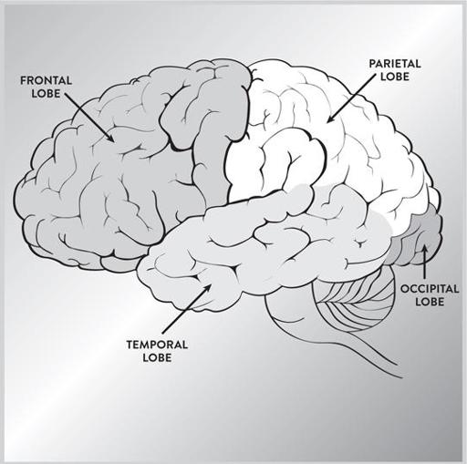
Figure 2
. The four lobes of the neocortex of the brain are responsible for different, though related, functions. (
illustration credit 1.2
)
The parietal lobe is located at the top of our brains. The right hemisphere controls sensory attention and body image; the left hemisphere controls skilled movements and some aspects of language. Damage to this area can cause many problems, such as difficulty in locating parts of your own body.
The occipital lobe is located at the very back of the brain and processes visual information from the eyes. Damage to this area can cause blindness and visual impairment.
The temporal lobe controls language (on the left side only), as well as the visual recognition of faces and certain emotional feelings. Damage to this lobe can leave us speechless or without the ability to recognize familiar faces.
THE EVOLVING BRAIN
When you look at other organs of the body, such as our muscles, bones, and lungs, there seems to be an obvious rhyme and reason to them that we can immediately see. But the structure of the brain might seem slapped together in a rather chaotic fashion. In fact, trying to map the brain has often been called “cartography for fools.”
To make sense of the seemingly random structure of the brain, in 1967 Dr. Paul MacLean of the National Institute of Mental Health applied Charles Darwin’s theory of evolution to the brain. He divided the brain into three parts. (Since then, studies have shown that there are refinements to this model, but we will use it as a rough organizing principle to explain the overall structure of the brain.) First, he noticed that the back and center part of our brains, containing the brain stem, cerebellum, and basal ganglia, are almost identical to the brains of reptiles. Known as the “reptilian brain,” these are the oldest structures of the brain, governing basic animal functions such as balance, breathing, digestion, heartbeat, and blood pressure. They also control behaviors such as fighting, hunting, mating, and territoriality, which are necessary for survival and reproduction. The reptilian brain can be traced back about 500 million years. (See
Figure 3
.)
But as we evolved from reptiles to mammals, the brain also became more complex, evolving outward and creating entirely new structures. Here we
encounter the “mammalian brain,” or the limbic system, which is located near the center of the brain, surrounding parts of the reptilian brain. The limbic system is prominent among animals living in social groups, such as the apes. It also contains structures that are involved in emotions. Since the dynamics of social groups can be quite complex, the limbic system is essential in sorting out potential enemies, allies, and rivals.