Read The Future of the Mind Online
Authors: Michio Kaku
The Future of the Mind (6 page)
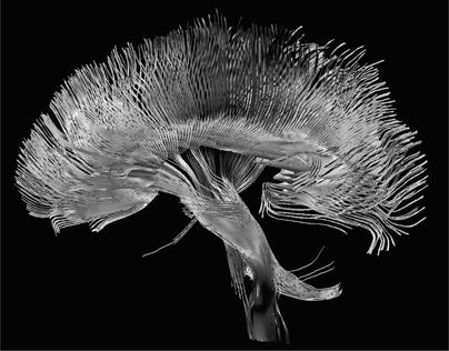
Figure 5
. At the top, we see an image taken by a functional MRI machine, showing regions of high mental activity. In the bottom image, we see the flowerlike pattern created by a diffusion MRI machine, which can follow the neural pathways and connections of the brain. (
illustration credit 1.5a
)
An EEG scan differs from an MRI scan in several crucial ways. The MRI scan, as we have seen, shoots radio pulses into the brain and then analyzes the “echoes” that come back. This means you can vary the radio pulse to select different atoms for analysis, making it quite versatile. The EEG machine, however, is strictly passive; that is, it analyzes the tiny electromagnetic signals the brain naturally emits. The EEG excels at recording the broad electromagnetic signals that surge across the entire brain, which allows scientists to measure the overall activity of the brain as it sleeps, concentrates, relaxes, dreams, etc. Different states of consciousness vibrate at different frequencies. For example, deep sleep corresponds to delta waves, which vibrate at .1 to 4 cycles per second. Active mental states, such as problem solving, correspond to beta waves, vibrating from 12 to 30 cycles per second. These vibrations allow various parts of the brain to share information and communicate with one another, even if they are located on opposite sides of the brain. And while MRI scans measuring blood flow can be taken only several times a second, EEG scans measure electrical activity instantly.
The greatest advantage of the EEG scan, though, is its convenience and cost. Even high school students have done experiments in their living rooms with EEG sensors placed over their heads.
However, the main drawback to the EEG, which has held up its development for decades, is its very poor spatial resolution. The EEG picks up electrical signals that have already been diffused after passing through the skull, making it difficult to detect abnormal activity when it originates deep in the brain. Looking at the output of the muddled EEG signals, it is almost impossible to say for sure which part of the brain created it. Furthermore, slight motions, like moving a finger, can distort the signal, sometimes rendering it useless.
PET SCANS
Yet another useful tool from the world of physics is the positron emission topography (PET) scan, which calculates the flow of energy in the brain by locating the presence of glucose, the sugar molecule that fuels cells. Like the cloud chamber I made as a high school student, PET scans make use of the subatomic particles emitted from sodium-22 within the glucose. To start the PET scan, a special solution containing slightly radioactive sugar is injected into the patient. The sodium atoms inside the sugar molecules have
been replaced by radioactive sodium-22 atoms. Every time a sodium atom decays, it emits a positive electron, or positron, which is easily detected by sensors. By following the path of the radioactive sodium atoms in sugar, one can then trace out the energy flow within the living brain.
The PET scan shares many of the same advantages of MRI scans but does not have the fine spatial resolution of an MRI photo. However, instead of measuring blood flow, which is only an indirect indicator of energy consumption in the body, PET scans measure energy consumption, so it is more closely related to neural activity.
There is another drawback to PET scans, however. Unlike MRI and EEG scans, PET scans are slightly radioactive, so patients cannot continually take them. In general, a person is not allowed to have a PET scan more than once a year because of the risk from radiation.
MAGNETISM IN THE BRAIN
Within the last decade, many new high-tech devices have entered the tool kit of neuroscientists, including the transcranial electromagnetic scanner (TES), magnetoencephalography (MEG), near-infrared spectroscopy (NIRS), and optogenetics, among others.
In particular, magnetism has been used to systematically shut down specific parts of the brain without cutting it open. The basic physics behind these new tools is that a rapidly changing electric field can create a magnetic field, and vice versa. MEGs passively measure the magnetic fields produced by the changing electric fields of the brain. These magnetic fields are weak and extremely tiny, only a billionth of Earth’s magnetic field. Like the EEG, the MEG is extremely good at time resolution, down to a thousandth of a second. Its spatial resolution, however, is only a cubic centimeter.
Unlike the passive measurement of the MEG, the TES generates a large pulse of electricity, which in turn creates a burst of magnetic energy. The TES is placed next to the brain, so the magnetic pulse penetrates the skull and creates yet another electric pulse inside the brain. This secondary electrical pulse, in turn, is sufficient to turn off or dampen the activity of selected areas of the brain.
Historically, scientists had to rely on strokes or tumors to silence certain parts of the brain and hence determine what they do. But with the TES, one can harmlessly turn off or dampen parts of the brain at will. By shooting
magnetic energy at a particular spot in the brain, one can determine its function by simply watching how a person’s behavior has changed. (For example, by shooting magnetic pulses into the left temporal lobe, one can see that this adversely affects our ability to talk.)
One potential drawback of the TES is that these magnetic fields do not penetrate very far into the interior of the brain (because magnetic fields decrease much faster than the usual inverse square law for electricity). TES is quite useful in turning off parts of the brain near the skull, but the magnetic
field cannot reach important centers located deep in the brain, such as the limbic system. But future generations of TES devices may overcome this technical problem by increasing the intensity and precision of the magnetic field.
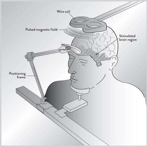
Figure 6
. We see the transcranial electromagnetic scanner and the magnetoencephalograph, which uses magnetism rather than radio waves to penetrate the skull and determine the nature of thoughts within the brain. Magnetism can temporarily silence parts of the brain, allowing scientists to safely determine how these regions perform without relying on stroke victims. (
illustration credit 1.6
)
DEEP BRAIN STIMULATION
Yet another tool that has proven vital to neurologists is deep brain stimulation (DBS). The probes originally used by Dr. Penfield were relatively crude. Today these electrodes can be hairlike and reach specific areas of the brain deep within its interior. Not only has DBS allowed scientists to locate the function of various parts of the brain, it can also be used to treat mental disorders. DBS has already proven its worth with Parkinson’s disease, in which certain regions of the brain are overactive and often create uncontrollable shaking of the hands.
More recently, these electrodes have targeted a new area of the brain (called Brodmann’s area number 25) that is often overactive in depressed patients who do not respond to psychotherapy or drugs. Deep brain stimulation has given almost miraculous relief after decades of torment and agony for these long-suffering patients.
Every year, new uses for deep brain stimulation are being found. In fact, nearly all the major disorders of the brain are being reexamined in light of this and other new brain-scanning technologies. This promises to be an exciting new area for diagnosing and even treating illnesses.
OPTOGENETICS—LIGHTING UP THE BRAIN
But perhaps the newest and most exciting instrument in the neurologist’s tool kit is optogenetics, which was once considered science fiction. Like a magic wand, it allows you to activate certain pathways controlling behavior by shining a light beam on the brain.
Incredibly, a light-sensitive gene that causes a cell to fire can be inserted, with surgical precision, directly into a neuron. Then, by turning on a light beam, the neuron is activated. More importantly, this allows scientists to excite these pathways, so that you can turn on and off certain behaviors by flicking a switch.
Although this technology is only a decade old, optogenetics has already proven successful in controlling certain animal behaviors. By turning on a light switch, it is possible to make fruit flies suddenly fly off, worms stop wiggling, and mice run around madly in circles. Monkey trials are now beginning, and even human trials are in discussion. There is great hope that this technology will have a direct application in treating disorders like Parkinson’s and depression.
THE TRANSPARENT BRAIN
Like optogenetics, another spectacular new development is making the brain fully transparent so that its neural pathways are exposed to the naked eye. In 2013, scientists at Stanford University announced that they had successfully made the entire brain of a mouse transparent, as well as parts of a human brain. The announcement was so stunning that it made the front page of the
New York Times
, with the headline “Brain as Clear as Jell-O for Scientists to Explore.”
At the cellular level, cells seen individually are transparent, with all their microscopic components fully exposed. However, once billions of cells come together to form organs like the brain, the addition of lipids (fats, oils, waxes, and chemicals not soluble in water) helps make the organ opaque. The key to the new technique is to remove the lipids while keeping the neurons intact. The scientists at Stanford did this by placing the brain in hydrogel (a gel-like substance mainly made of water), which binds to all the brain’s molecules except the lipids. By placing the brain in a soapy solution with an electric field, the solution can be flushed out of the brain, carrying along the lipids, leaving the brain transparent. The addition of dyes can then make the neural pathways visible. This will help to identify and map the many neural pathways of the brain.
Making tissue transparent is not new, but getting precisely the right conditions necessary to make the entire brain transparent took a lot of ingenuity. “I burned and melted more than a hundred brains,” confessed Dr. Kwanghun Chung, one of the lead scientists in the study. The new technique, called Clarity, can also be applied to other organs (and even organs preserved years ago in chemicals like formalin). He has already created transparent livers, lungs, and hearts. This new technique has startling applications across all of
medicine. In particular, it will accelerate locating the neural pathways of the brain, which is the focus of intense research and funding.
FOUR FUNDAMENTAL FORCES
The success of this first generation of brain scans has been nothing less than spectacular. Before their introduction, only about thirty or so regions of the brain were known with any certainty. Now the MRI machine alone can identify two to three hundred regions of the brain, opening up entirely new frontiers for brain science. With so many new scanning technologies being introduced from physics just within the last fifteen years, one might wonder: Are there more? The answer is yes, but they will be variations and refinements of the previous ones, not radically new technologies. This is because there are only four fundamental forces—gravitational, electromagnetic, weak nuclear, and strong nuclear—that rule the universe. (Physicists have tried to find evidence for a fifth force, but so far all such attempts have failed.)
The electromagnetic force, which lights up our cities and represents the energy of electricity and magnetism, is the source of almost all the new scanning technologies (with the exception of the PET scan, which is governed by the weak nuclear force). Because physicists have had over 150 years of experience working with the electromagnetic force, there is no mystery in creating new electric and magnetic fields, so any new brain-scanning technology will most likely be a novel modification of existing technologies, rather than being something entirely new. As with most technology, the size and cost of these machines will drop, vastly increasing the widespread use of these sophisticated instruments. Already physicists are doing the basic calculations necessary to make an MRI machine fit into a cell phone. At the same time, the fundamental challenge facing these brain scans is resolution, both spatial and temporal. The spatial resolution of MRI scans will increase as the magnetic field becomes more uniform and as the electronics become more sensitive. At present, MRI scans can see only dots or voxels within a fraction of a millimeter. But each dot may contain hundreds of thousands of neurons. New scanning technology should reduce this even further. The holy grail of this approach would be to create an MRI-like machine that could identify individual neurons and their connections.