Read The Future of the Mind Online
Authors: Michio Kaku
The Future of the Mind (5 page)
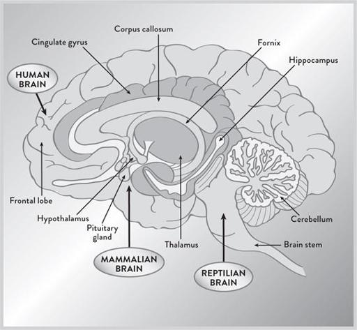
Figure 3
. The evolutionary history of the brain, with the reptilian brain, the limbic system (the mammalian brain), and the neocortex (the human brain). Roughly speaking, one can argue that the path of our brain’s evolution passed from the reptilian brain to the mammalian brain to the human brain. (
illustration credit 1.3
)
The different parts of the limbic system that control behaviors crucial for social animals are:
• The hippocampus. This is the gateway to memory, where short-term memories are processed into long-term memories. Its name means
“seahorse,” which describes its strange shape. Damage here will destroy the ability to make new long-term memories. You are left a prisoner of the present.
• The amygdala. This is the seat of emotions, especially fear, where emotions are first registered and generated. Its name means “almond.”
• The thalamus. This is like a relay station, gathering sensory signals from the brain stem and then sending them out to the various cortices. Its name means “inner chamber.”
• The hypothalamus. This regulates body temperature, our circadian rhythm, hunger, thirst, and aspects of reproduction and pleasure. It lies below the thalamus—hence its name.
Finally, we have the third and most recent region of the mammalian brain, the cerebral cortex, which is the outer layer of the brain. The latest evolutionary structure within the cerebral cortex is the neocortex (meaning “new bark”), which governs higher cognitive behavior. It is most highly developed in humans: it makes up 80 percent of our brain’s mass, yet is only as thick as a napkin. In rats the neocortex is smooth, but it is highly convoluted in humans, which allows a large amount of surface area to be crammed into the human skull.
In some sense, the human brain is like a museum containing remnants of all the previous stages in our evolution over millions of years, exploding outward and forward in size and function. (This is also roughly the path taken when an infant is born. The infant brain expands outward and toward the front, perhaps mimicking the stages of our evolution.)
Although the neocortex seems unassuming, looks are deceiving. Under a microscope you can appreciate the intricate architecture of the brain. The gray matter of the brain consists of billions of tiny brain cells called neurons. Like a gigantic telephone network, they receive messages from other neurons via dendrites, which are like tendrils sprouting from one end of the neuron. At the other end of the neuron, there is a long fiber called the axon. Eventually the axon connects to as many as ten thousand other neurons via their dendrites. At the juncture between the two, there is a tiny gap called the synapse. These synapses act like gates, regulating the flow of information within the brain. Special chemicals called neurotransmitters can enter the synapse and alter the flow of signals. Because neurotransmitters like dopamine, serotonin,
and noradrenaline help control the stream of information moving across the myriad pathways of the brain, they exert a powerful effect on our moods, emotions, thoughts, and state of mind. (See
Figure 4
.)
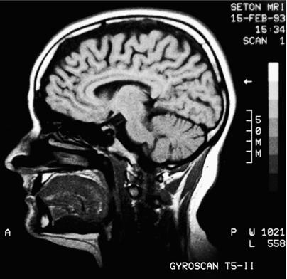
This description of the brain roughly represented the state of knowledge through the 1980s. In the 1990s, however, with the introduction of new technologies from the field of physics, the mechanics of thought began to be revealed in exquisite detail, unleashing the current explosion of scientific discovery. One of the workhorses of this revolution has been the MRI machine.
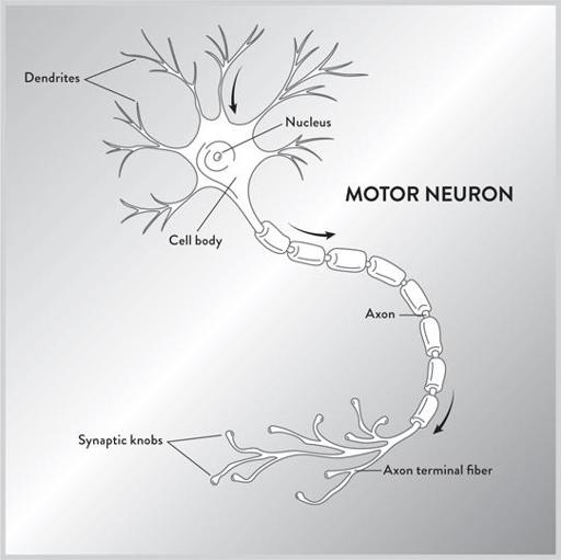
Figure 4
. Diagram of a neuron. Electrical signals travel along the axon of the neuron until they hit the synapse. Neurotransmitters can regulate the flow of electrical signals past the synapse. (
illustration credit 1.4
)
THE MRI: WINDOW INTO THE BRAIN
To understand the reason why this radical new technology has helped decode the thinking brain, we have to turn our attention to some basic principles of physics.
Radio waves, a type of electromagnetic radiation, can pass right through tissue without doing damage. MRI machines take advantage of this fact, allowing electromagnetic waves to freely penetrate the skull. In the process, this technology has given us glorious photographs of something once thought to be impossible to capture: the inner workings of the brain as it experiences sensations and emotions. Watching the dance of lights flickering in a MRI machine, one can trace out the thoughts moving within the brain. It’s like being able to see the inside of a clock as it ticks.
The first thing you notice about an MRI machine is the huge, cylindrical magnetic coils, which can produce a magnetic field twenty to sixty thousand times greater than the strength of Earth’s. The giant magnet is one of the principal reasons why an MRI machine can weigh a ton, fill up an entire room, and cost several million dollars. (MRI machines are safer than X-ray machines because they don’t create harmful ions. CT scans, which can also create 3-D pictures, flood the body with many times the dosage from an ordinary X-ray, and hence have to be carefully regulated. By contrast, MRI machines are safe when used properly. One problem, however, is the carelessness of workers. The magnetic field is powerful enough to send tools hurling through the air at high velocity when turned on at the wrong time. People have been injured and even killed in this way.)
MRI machines work as follows: Patients lie flat and are inserted into a cylinder containing two large coils, which create the magnetic field. When the magnetic field is turned on, the nuclei of the atoms inside your body act very much like a compass needle: they align horizontally along the direction of the field. Then a small pulse of radio energy is generated, which causes some of the nuclei in our body to flip upside down. When the nuclei later revert back to their normal position, they emit a secondary pulse of radio energy, which is then analyzed by the MRI machine. By analyzing these tiny “echoes,” one can then reconstruct the location and nature of these atoms. Like a bat, which uses echoes to determine the position of objects in its path, the echoes created by the MRI machine allow scientists to re-create
a remarkable image of the inside of the brain. Computers then reconstruct the position of the atoms, giving us beautiful diagrams in three dimensions.
When MRIs were originally introduced, they were able to show the static structure of the brain and its various regions. However, in the mid-1990s, a new type of MRI was invented, called “functional” MRI, or fMRI, which detected the presence of oxygen in the blood in the brain. (For different types of MRI machines, scientists sometimes put a lowercase letter in front of “MRI,” but we will use the abbreviation MRI to denote all the various types of MRI machines.) MRI scans cannot directly detect the flow of electricity in the neurons, but since oxygen is necessary to provide the energy for the neurons, oxygenated blood can indirectly trace the flow of electrical energy in the neurons and show how various regions of the brain interact with one another.
Already these MRI scans have definitively disproven the idea that thinking is concentrated in a single center. Instead, one can see electrical energy circulating across different parts of the brain as it thinks. By tracing the path taken by our thoughts, MRI scans have shed new light into the nature of Alzheimer’s, Parkinson’s, schizophrenia, and a host of other mental diseases.
The great advantage of MRI machines is their exquisite ability to locate minute parts of the brain, down to a fraction of a millimeter in size. An MRI scan will create not just dots on a two-dimensional screen, called pixels, but dots in three-dimensional space, called “voxels,” yielding a bright collection of tens of thousands of colored dots in 3-D, in the shape of a brain.
Since different chemical elements respond to different frequencies of radio, you can change the frequency of the radio pulse and therefore identify different elements of the body. As noted, fMRI machines zero in on the oxygen atom contained within blood in order to measure blood flow, but MRI machines can also be tuned to identify other atoms. In just the last decade, a new form of MRI was introduced called “diffusion tensor imaging” MRI, which detects the flow of water in the brain. Since water follows the neural pathways of the brain, DTI yields beautiful pictures that resemble networks of vines growing in a garden. Scientists can now instantly determine how certain parts of the brain are hooked up with other parts.
There are a couple of drawbacks to MRI technology, however. Although they are unparalleled in spatial resolution, locating voxels down to the size of a pinpoint in three dimensions, MRIs are not that good in temporal resolution.
It takes almost a full second to follow the path of blood in the brain, which may not sound like a lot, but remember that electrical signals travel almost instantly throughout the brain, and hence MRI scans can miss some of the intricate details of thought patterns.
Another snag is the cost, which runs in the millions of dollars, so doctors often have to share the machines. But like most technology, developments should bring down the cost over time.
In the meantime, exorbitant costs haven’t stalled the hunt for commercial applications. One idea is to use MRI scans as lie detectors, which, according to some studies, can identify lies with 95 percent accuracy or higher. The level of accuracy is still controversial, but the basic idea is that when a person tells a lie, he simultaneously has to know the truth, concoct the lie, and rapidly analyze the consistency of this lie with previously known facts. Today some companies are claiming that MRI technology shows that the prefrontal and parietal lobes light up when someone tells a lie. More specifically, the “orbitofrontal cortex” (which can serve, among other functions, as the brain’s “fact-checker” to warn us when something is wrong) becomes active. This area is located right behind the orbits of our eyes, and hence the name. The theory goes that the orbitofrontal cortex understands the difference between the truth and a lie and kicks into overdrive as a result. (Other areas of the brain also light up when someone tells a lie, such as the superiormedial and inferolateral prefrontal cortices, which are involved in cognition.)
Already there are several commercial firms offering MRI machines as lie detectors, and cases involving these machines are entering the court system. But it’s important to note that these MRI scans indicate increased brain activity only in certain areas. While DNA results can sometimes have an accuracy of one part in 10 billion or better, MRI scans cannot, because it takes many areas of the brain to concoct a lie, and these same areas of the brain are responsible for processing other kinds of thoughts as well.
EEG SCANS
Another useful tool to probe deep inside the brain is the EEG, the electroencephalogram. The EEG was introduced all the way back in 1924, but only recently has it been possible to employ computers to make sense out of all the data pouring in from each electrode.
To use the EEG machine, the patient usually puts on a futuristic-looking helmet with scores of electrodes on the surface. (More advanced versions place a hairnet over the head containing a series of tiny electrodes.) These electrodes detect the tiny electrical signals that are circulating in the brain.